
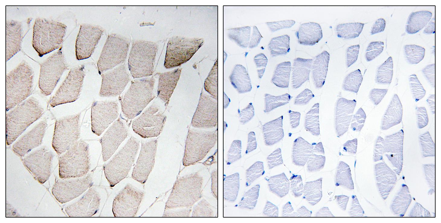
Target:Myomesin-1
Gene Name:MYOM1
Protein Name:Myomesin-1
Human Gene Id:8736
Human Swiss Prot No:P52179
Mouse Swiss Prot No:Q62234
Immunogen:The antiserum was produced against synthesized peptide derived from human MYOM1. AA range:824-873
Specificity:Myomesin-1 Polyclonal Antibody detects endogenous levels of Myomesin-1 protein.
Formulation:Liquid in PBS containing 50% glycerol, 0.5% BSA and 0.02% sodium azide.
Source:Polyclonal, Rabbit,IgG
Dilution:IHC 1:100 - 1:300. ELISA: 1:40000.. IF 1:50-200
Purification:The antibody was affinity-purified from rabbit antiserum by affinity-chromatography using epitope-specific immunogen.
Concentration:1 mg/ml
Storage Stability:-15°C to -25°C/1 year(Do not lower than -25°C)
Other Name:MYOM1;Myomesin-1;190 kDa connectin-associated protein;190 kDa titin-associated protein;Myomesin family member 1
Molecular Weight(Da):162kD
Background: The giant protein titin, together with its associated proteins, interconnects the major structure of sarcomeres, the M bands and Z discs. The C-terminal end of the titin string extends into the M line, where it binds tightly to M-band constituents of apparent molecular masses of 190 kD (myomesin 1) and 165 kD (myomesin 2). This protein, myomesin 1, like myomesin 2, titin, and other myofibrillar proteins contains structural modules with strong homology to either fibronectin type III (motif I) or immunoglobulin C2 (motif II) domains. Myomesin 1 and myomesin 2 each have a unique N-terminal region followed by 12 modules of motif I or motif II, in the arrangement II-II-I-I-I-I-I-II-II-II-II-II. The two proteins share 50% sequence identity in this repeat-containing region. The head structure formed by these 2 proteins on one end of the titin string extends into the center of the M band. The integrating structure
Function:function:Major component of the vertebrate myofibrillar M band. Binds myosin, titin, and light meromyosin. This binding is dose dependent.,similarity:Contains 5 fibronectin type-III domains.,similarity:Contains 5 Ig-like C2-type (immunoglobulin-like) domains.,subunit:Interacts with TTN/titin (By similarity). Interacts with PNKD.,
Subcellular Location:Cytoplasm, myofibril, sarcomere, M line .
Expression: Heart muscle,Skeletal muscle,
商品信息已成功复制,启研竭诚为您服务


