
Target:PEA-15
Gene Name:PEA15
Protein Name:Astrocytic phosphoprotein PEA-15
Human Gene Id:8682
Human Swiss Prot No:Q15121
Mouse Gene Id:18611
Mouse Swiss Prot No:Q62048
Rat Gene Id:364052
Rat Swiss Prot No:Q5U318
Immunogen:The antiserum was produced against synthesized peptide derived from human PEA-15. AA range:81-130
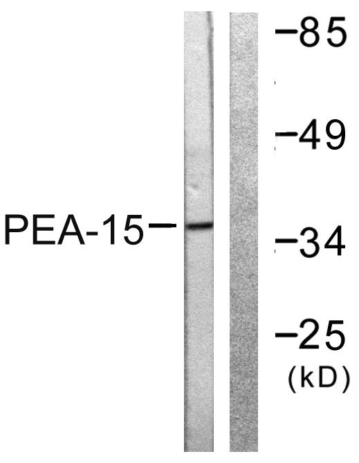
Specificity:PEA-15 Polyclonal Antibody detects endogenous levels of PEA-15 protein.
Formulation:Liquid in PBS containing 50% glycerol, 0.5% BSA and 0.02% sodium azide.
Source:Polyclonal, Rabbit,IgG
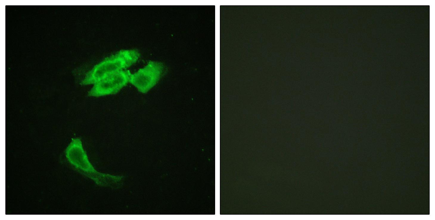
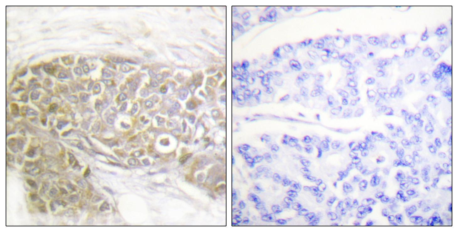
Dilution:WB 1:500 - 1:2000. IHC 1:100 - 1:300. IF 1:200 - 1:1000. ELISA: 1:40000. Not yet tested in other applications.
Purification:The antibody was affinity-purified from rabbit antiserum by affinity-chromatography using epitope-specific immunogen.
Concentration:1 mg/ml
Storage Stability:-15°C to -25°C/1 year(Do not lower than -25°C)
Other Name:PEA15;Astrocytic phosphoprotein PEA-15;15 kDa phosphoprotein enriched in astrocytes;Phosphoprotein enriched in diabetes;PED
Observed Band(KD):36kD
Background:phosphoprotein enriched in astrocytes 15(PEA15) Homo sapiens This gene encodes a death effector domain-containing protein that functions as a negative regulator of apoptosis. The encoded protein is an endogenous substrate for protein kinase C. This protein is also overexpressed in type 2 diabetes mellitus, where it may contribute to insulin resistance in glucose uptake. Alternative splicing results in multiple transcript variants. [provided by RefSeq, Jul 2014],
Function:function:Blocks Ras-mediated inhibition of integrin activation and modulates the ERK MAP kinase cascade. Inhibits RPS6KA3 activities by retaining it in the cytoplasm (By similarity). Inhibits both TNFRSF6- and TNFRSF1A-mediated CASP8 activity and apoptosis. Regulates glucose transport by controlling both the content of SLC2A1 glucose transporters on the plasma membrane and the insulin-dependent trafficking of SLC2A4 from the cell interior to the surface.,PTM:Phosphorylated by protein kinase C and calcium-calmodulin-dependent protein kinase. These phosphorylation events are modulated by neurotransmitters or hormones.,similarity:Contains 1 DED (death effector) domain.,subcellular location:Associated with microtubules.,subunit:Binds RPS6KA3, MAPK3 and MAPK1. Transient interaction with PLD1 and PLD2 (By similarity). Interacts with CASP8 and FADD.,tissue specificity:Ubiquitously expressed. Mo
Subcellular Location:Cytoplasm. Associated with microtubules.
Expression:Ubiquitously expressed. Most abundant in tissues such as heart, brain, muscle and adipose tissue which utilize glucose as an energy source. Lower expression in glucose-producing tissues. Higher levels of expression are found in tissues from individuals with type 2 diabetes than in controls.
商品信息已成功复制,启研竭诚为您服务


