
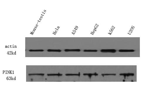
Target:PINK1
Fields:Mitophagy - animal;Parkinson disease;Amyotrophic lateral sclerosis;Pathways of neurodegeneration - multiple diseases
Gene Name:PINK1
Protein Name:Serine/threonine-protein kinase PINK1, mitochondrial (EC 2.7.11.1) (BRPK) (PTEN-induced putative kinase protein 1)
Human Gene Id:65018
Human Swiss Prot No:Q9BXM7
Mouse Swiss Prot No:Q99MQ3
Immunogen:Synthesized peptide derived from part region of human protein
Specificity:PINK1 Polyclonal Antibody detects endogenous levels of protein.
Formulation:Liquid in PBS containing 50% glycerol, and 0.02% sodium azide.
Source:Polyclonal, Rabbit,IgG
Dilution:WB 1:500-2000 ELISA 1:5000-20000
Purification:The antibody was affinity-purified from rabbit antiserum by affinity-chromatography using epitope-specific immunogen.
Concentration:1 mg/ml
Storage Stability:-15°C to -25°C/1 year(Do not lower than -25°C)
Observed Band(KD):63kD
Background: This gene encodes a serine/threonine protein kinase that localizes to mitochondria. It is thought to protect cells from stress-induced mitochondrial dysfunction. Mutations in this gene cause one form of autosomal recessive early-onset Parkinson disease. [provided by RefSeq, Jul 2008],
Function:catalytic activity:ATP + a protein = ADP + a phosphoprotein.,cofactor:Magnesium.,disease:Defects in PINK1 are the cause of autosomal recessive early-onset Parkinson disease 6 (PARK6) [MIM:605909, 168600]. Parkinson disease (PD) is a complex, multifactorial disorder that typically manifests after the age of 50 years, although early-onset cases (before 50 years) are known. PD generally arises as a sporadic condition but is occasionally inherited as a simple mendelian trait. Although sporadic and familial PD are very similar, inherited forms of the disease usually begin at earlier ages and are associated with atypical clinical features. PD is characterized by bradykinesia, resting tremor, muscular rigidity and postural instability, as well as by a clinically significant response to treatment with levodopa. The pathology involves the loss of dopaminergic neurons in the substantia nigra and t
Subcellular Location:Mitochondrion outer membrane ; Single-pass membrane protein . Mitochondrion inner membrane ; Single-pass membrane protein . Cytoplasm, cytosol . Localizes mostly in mitochondrion and the two smaller proteolytic processed fragments localize mainly in cytosol (PubMed:19229105). When mitochondria lose mitochondrial membrane potential following damage, PINK1 import is arrested, which induces its accumulation in the outer mitochondrial membrane, where it acquires kinase activity (PubMed:18957282). .
Expression:Highly expressed in heart, skeletal muscle and testis, and at lower levels in brain, placenta, liver, kidney, pancreas, prostate, ovary and small intestine. Present in the embryonic testis from an early stage of development.
商品信息已成功复制,启研竭诚为您服务


