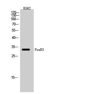
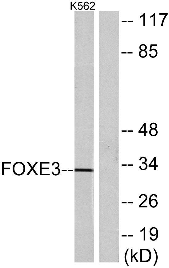
Target:FoxE3
Gene Name:FOXE3
Protein Name:Forkhead box protein E3
Human Gene Id:2301
Human Swiss Prot No:Q13461
Mouse Gene Id:30923
Mouse Swiss Prot No:Q9QY14
Rat Swiss Prot No:Q63250
Immunogen:The antiserum was produced against synthesized peptide derived from human FOXE3. AA range:81-130
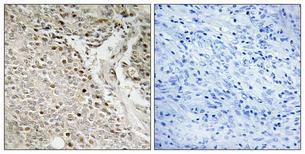
Specificity:FoxE3 Polyclonal Antibody detects endogenous levels of FoxE3 protein.
Formulation:Liquid in PBS containing 50% glycerol, 0.5% BSA and 0.02% sodium azide.
Source:Polyclonal, Rabbit,IgG
Dilution:WB 1:500 - 1:2000. IHC 1:100 - 1:300. ELISA: 1:20000.. IF 1:50-200
Purification:The antibody was affinity-purified from rabbit antiserum by affinity-chromatography using epitope-specific immunogen.
Concentration:1 mg/ml
Storage Stability:-15°C to -25°C/1 year(Do not lower than -25°C)
Other Name:FOXE3;FKHL12;FREAC8;Forkhead box protein E3;Forkhead-related protein FKHL12;Forkhead-related transcription factor 8;FREAC-8
Observed Band(KD):33kD
Background: This intronless gene belongs to the forkhead family of transcription factors, which is characterized by a distinct forkhead domain. The protein encoded functions as a lens-specific transcription factor and plays an important role in vertebrate lens formation. Mutations in this gene are associated with anterior segment mesenchymal dysgenesis and congenital primary aphakia. [provided by RefSeq, Dec 2009],
Function:disease:Defects in FOXE3 are a cause of anterior segment mesenchymal dysgenesis (ASMD) [MIM:107250]; also known as anterior segment ocular dysgenesis (ASOD). ASMD includes all malformations involving the first (corneal endothelium and trabecular meshwork), second (corneal stroma) and third (iris stroma) mesenchymal waves of neural crest. The ASMD phenotype is characterized by corneal opacities with or without iris adhesions in 100%, cataracts of varying severity in 100% and optic-nerve abnormalities in 20% of affected individuals.,disease:Defects in FOXE3 are a cause of congenital primary aphakia (CPA) [MIM:610256]. Human aphakia is a rare congenital eye disorder in which the lens is missing. It has been histologically subdivided into primary and secondary forms, in accordance with the severity of defects of the ocular tissues, whose development requires the initial presence of a lens. C
Subcellular Location:Nucleus .
商品信息已成功复制,启研竭诚为您服务