
Target:AMID
Fields:p53 signaling pathway
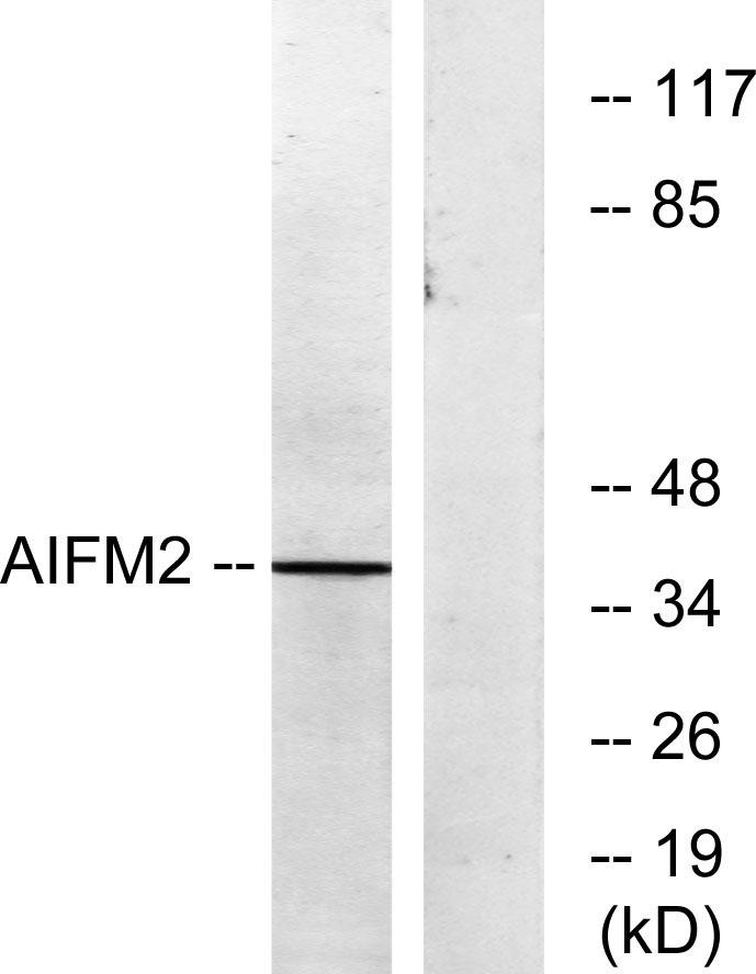
Gene Name:AIFM2
Protein Name:Apoptosis-inducing factor 2
Human Gene Id:84883
Human Swiss Prot No:Q9BRQ8
Mouse Gene Id:71361
Mouse Swiss Prot No:Q8BUE4
Immunogen:The antiserum was produced against synthesized peptide derived from human AIFM2. AA range:141-190
Specificity:AMID Polyclonal Antibody detects endogenous levels of AMID protein.
Formulation:Liquid in PBS containing 50% glycerol, 0.5% BSA and 0.02% sodium azide.
Source:Polyclonal, Rabbit,IgG
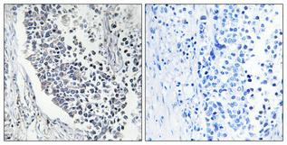
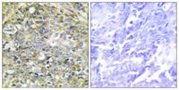
Dilution:WB 1:500 - 1:2000. IHC 1:100 - 1:300. IF 1:200 - 1:1000. ELISA: 1:40000. Not yet tested in other applications.
Purification:The antibody was affinity-purified from rabbit antiserum by affinity-chromatography using epitope-specific immunogen.
Concentration:1 mg/ml
Storage Stability:-15°C to -25°C/1 year(Do not lower than -25°C)
Other Name:AIFM2;AMID;PRG3;Apoptosis-inducing factor 2;Apoptosis-inducing factor homologous mitochondrion-associated inducer of death;Apoptosis-inducing factor-like mitochondrion-associated inducer of death;p53-responsive gene 3 protein
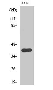
Observed Band(KD):41kD
Background: This gene encodes a flavoprotein oxidoreductase that binds single stranded DNA and is thought to contribute to apoptosis in the presence of bacterial and viral DNA. The expression of this gene is also found to be induced by tumor suppressor protein p53 in colon cancer cells. [provided by RefSeq, Nov 2010],
Function:cofactor:FAD. Binds 6-hydroxy-FAD non-covalently.,function:Oxidoreductase, which may play a role in mediating a TP53/p53-dependent apoptosis response. Probable oxidoreductase that acts as a caspase-independent mitochondrial effector of apoptotic cell death. Binds to DNA in a sequence-independent manner. May contribute to genotoxin-induced growth arrest.,induction:Expression detected at 4 hours after induction by TP53/p53. Down-regulated in a wide range of human tumors.,similarity:Belongs to the FAD-dependent oxidoreductase family.,tissue specificity:Detected in most normal tissues as two transcripts of 1.8 and 4.0 kb in length, respectively. Highly expressed in heart, moderately in liver and skeletal muscles, and expressed at low levels in placenta, lung, kidney, and pancreas. Both transcripts expressed following TP53/p53 induction. The shorter 1.8 kb transcript seems to be the major tra
Subcellular Location:Lipid droplet . Cell membrane ; Lipid-anchor . Cytoplasm . Mitochondrion membrane . Nucleus .
Expression:Detected in most normal tissues as two transcripts of 1.8 and 4.0 kb in length, respectively. Highly expressed in heart, moderately in liver and skeletal muscles, and expressed at low levels in placenta, lung, kidney, and pancreas. Both transcripts expressed following p53/TP53 induction. The shorter 1.8 kb transcript seems to be the major transcript in EB1 colon cancer cells.
商品信息已成功复制,启研竭诚为您服务


